Cell culture It is one of the most important tools used in cellular and molecular biology, as it provides excellent model systems for studying normal cell physiology and biochemistry (e.g. metabolic studies, aging), the effects of drugs and toxic compounds on cells, mutagenesis and carcinogenesis. It is also used in drug screening and development and in the large-scale manufacture of biological compounds (e.g. vaccines, therapeutic proteins). The main advantage of using cell culture for any of these applications is the consistency and reproducibility of results that can be obtained by using a batch of clonable cells.
Cell culture refers to the removal of cells from an animal or plant and their subsequent growth in a favorable artificial environment. Cells may be removed from the tissue directly and broken down by enzymatic or mechanical means prior to culture, or they may be derived from a cell line or cell strain that has already been established.
Primary culture
Primary culture refers to the stage of culture after cells are isolated from the tissue and proliferated under appropriate conditions until they occupy all available substrate (i.e., reach confluence). At this stage, cells must be subcultured (i.e., passaged) by transferring them to a new vessel with fresh growth medium to provide more space for continued growth.
Cell line
After the first subculture, the primary culture is referred to as a cell line or sub-clone. Cell lines derived from primary cultures have a limited lifespan (i.e., they are finite) and as they are passaged, cells with the greatest growth capacity predominate, resulting in a degree of genotypic and phenotypic uniformity in the population.
Cell strain
If a subpopulation of a cell line is positively selected from the culture through cloning or some other method, this cell line becomes a cell strain. A cell strain often acquires additional genetic changes subsequent to the initiation of the parental line.
Finite vs. continuous cell lines
Normal cells typically divide a limited number of times before losing their ability to proliferate, an event genetically known as senescence. These cell lines are known as finite. However, some cell lines become immortal through a process called transformation, which can occur spontaneously or can be induced chemically or virally. When a finite cell line is transformed and gains the ability to divide indefinitely, it becomes a continuous cell line.
Cell lines in continuous culture are prone to genetic drift. Finite cell lines are destined for senescence. All cell cultures are susceptible to microbial contamination, and even the best laboratories can experience equipment failure. Because an established cell line is a valuable resource and is expensive and time-consuming to replace, it is vitally important that they be frozen and preserved for long-term storage.
As soon as a small surplus of subculture cells is available, they should be frozen as seed stock, protected, and not made available for general laboratory use. Working stock can be prepared and replenished from frozen seed stocks. If seed stocks become depleted, cryopreserved working stocks can serve as a source for preparing a fresh seed stock with minimal increase in generation number since initial freezing.
The best method for cryopreserving cultured cells is to store them in liquid nitrogen in complete medium in the presence of a cryoprotective agent such as dimethyl sulfoxide (DMSO) or glycerol. Cryoprotective agents lower the freezing point of the medium and also allow for a slower cooling rate, which greatly reduces the risk of ice crystal formation, which can damage cells and cause cell death.
DMSO is known to facilitate the entry of organic molecules into tissues. Handle reagents containing DMSO using equipment and practices appropriate to the hazards posed by such materials. Dispose of reagents in accordance with local regulations.
Cell culture from a frozen cell stock
Once cells have reached approximately 90% confluence, cell stocks can be expanded using T150 flasks.
Make sure to complete the cell bank registration.
Determine the passage number, in. If the previous cells were frozen, passaged three times, the thawing process would be the fourth step.
Add growth media (Basic Media, Calf Serum, and Antibiotics) to a T75 flask labeled with the cell line, passage number, and date.
Place the flask horizontally in a CO2 incubator at 37°C and 5% CO2 for at least 15 minutes. This will allow the medium to warm up and reach its normal pH (7.0-7.6)
Thaw the frozen vial with the cell line by gentle agitation in a 37°C water bath. To reduce the risk of contamination, keep the O-ring and cap above the water level (2-5 minutes).
Place the vial in the biosafety cabinet (BSL-2). To avoid contamination, clean the vial with 70% ethanol before opening.
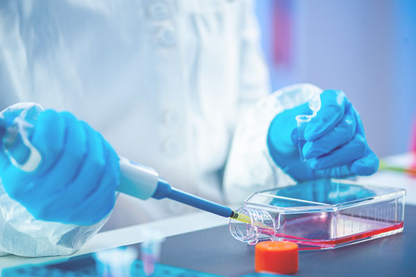
Transfer the contents of the frozen cell vial to the T75 flask containing growth media. Gently mix and swirl the flask to distribute the cells evenly across the surface of the flask.
Incubate the flask overnight in a CO2 incubator at 37°C, 5% CO2. Allow cells to attach overnight.
Change the growth media.
Incubate the flask at 37°C, 5% CO2 for an additional 2-4 days and monitor for cell growth.