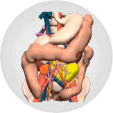
3D VIRTUAL AND PHYSICAL MODELS
In collaboration with Cella Medical Solutions, we perform high-precision 3D reconstructions of the patient’s anatomy for the planning of complex surgeries.
For surgeons, the use of 3D models represents a differential advantage, as it allows them to obtain better clinical results, with less risk for the patient and with significant savings in operating time and costs for the hospital.
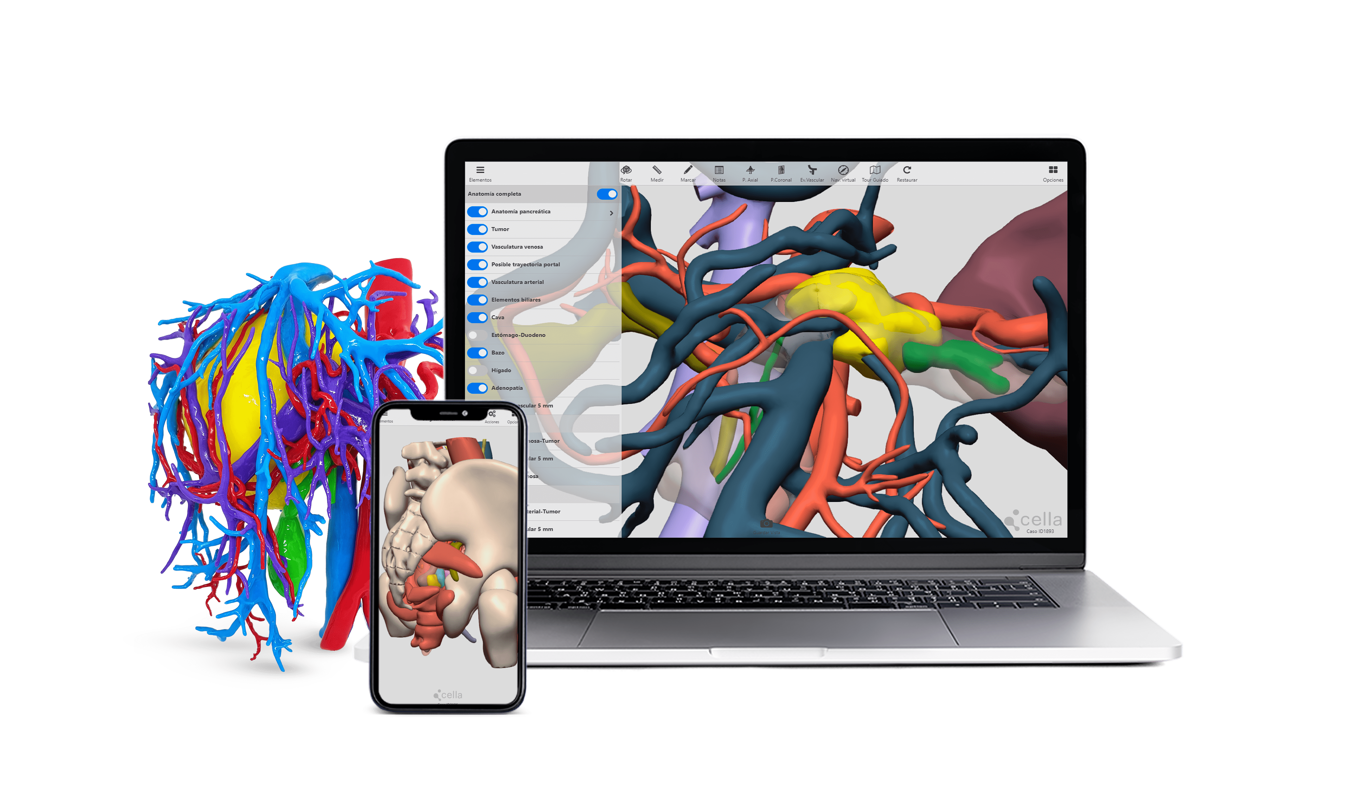
Access the 3D virtual models through our web platform from computers, tablets, smartphones, etc. The virtual models allow complete interaction with the patient’s anatomy and have a differential advantage for surgical planning, as they include tools and functions adapted to the needs of each specialty.
Access to the Cella platform

3D surgical planner
Features
- - Relevant medical information: vascular variants, hepatic segments, infiltrations, interactions...
- - Simulate surgical decisions: resection margins, fixed and fixed resections.
- - Rotate, zoom, hide and define the transparency of the different structures.
- - Check the distances, know your volumes...
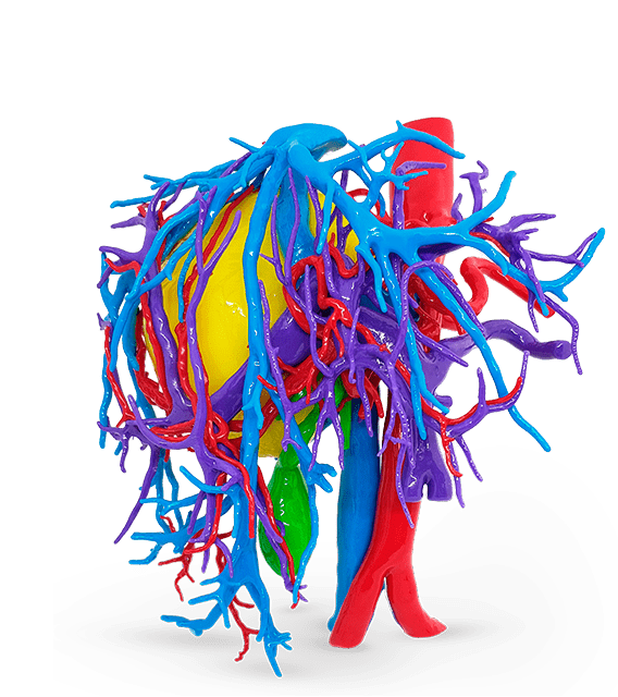
At CellaMedicalSolutions we make 3D printed reconstructions of the patient’s anatomy and body to assist surgeons in the planning of surgeries. Our 3D printed models are manufactured with a high degree of precision and quality. Each case goes through a study process to create models adapted to the surgical procedure, differentiating the structures by color and transparency.
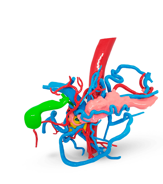
Features
- -Reconstruction at the same time and sterilizable by conventional methods.
- -Differentiation of structures through transparencies and multiple colors. Rigid material with emulated texture, which simulates the physical properties of real tissues (elasticity, cut, suture).
- -Simulation of surgeries.
- -High precision and quality.
- -Profitable models developed with proprietary technology.
The models are created with medical images of the patient (MRI, CT, PET, combination of these) through advanced algorithms, artificial intelligence and medical image processing that are supervised by specialized radiologists. The models are created with medical images of the patient (MRI, CT, PET, combination of these) through advanced algorithms, artificial intelligence and medical image processing that are supervised by specialized radiologists.

SURGICAL SPECIALITIES

Urological

Hepatic

Pancreatic
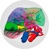
Biliary

Esophagogastric

Colon
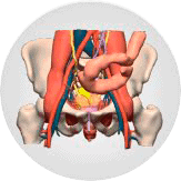
Rectum
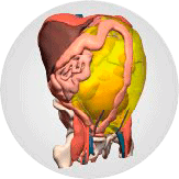
Peritoneal and retroperitoneal
